Tuesday, December 6, 2011

Descriptive Chart

Here are my final results!

MORPHOLOGICAL CHARACTERISTICS
Cell shape: Bacilli
Spores: No endospores
Gram's Stain: Gram negative
Motility: Not motile
Capsules: None
Special Stains: Non-acid fast

CULTURAL CHARACTERISTICS
Colonies
     Nutrient Agar: Did not do
     Blood Agar: Did not do
Agar Slant: White colony
Nutrient Broth: Growth throughout broth
Oxygen Requirements: Facultative
Optimum Temp: 37 degree's C


PHYSIOLOGICAL CHARACTERISTICS

TEST
RESULTS
FERMENTATION
Glucose
Acid / Gas
Lactose
Acid/ Gas
Sucrose
No Fermentation
Mannitol
Non-Fermenter
HYDROLYSIS
Gelatin Liquefaction
Negative
Starch
Positive
Casein
Negative
Fat
Negative
IMVIC
Indole
Positive
Methyl Red
Positive
Voges-Proskauer
Negative
Citrate Utilization
Negative
Nitrate Reduction
Positive
Urease
Negative
Catalase
Negative (very weak bubbling)
Oxidase
Negative


Because my bacteria is a Gram negative, Bacilli shaped, Lactose positive, Indole positive, and Citrate negative bacteria, My bacteria is.......E. Coli!

Monday, December 5, 2011

Nov 22

We did a an exercise studying the technique of immunology and we applied it in a test for food purity. Today I worked with Katherine.

What is immunology? = the Study of an organism's response to a foreign organic substance.

What we did
Took a dropping pipet and a petri dish containing agar. The bottom of each dish was labeled I, II, or III.

Placed the petri dish over the appropriate template so the template and section matched.
Held the dropping pipet bertically over one of the circles on teh template. Squeezing the pipet bulb we gently touched the pipet tip to the surface of the agar. While relasing the bulb we pushed the pipet tip down through the agar to the bottom of the dish, and lifted the pipet vertically. This left a straight- walled well in the agar.

Then, following the instructions, I filled each well.

EXERCISE 1 - Diffusion and Precipitation in Agar

Using template I, I made four wells and then filled them.

well 1 = Green Dye
well 2 = Red Dye
well 3 = Barium Chloride
well 4 = Potassium Sulfate

Closer look at wells and dye
Adding the dyes


Results

EXERCISE 2 - Anitbody-antigen Reaction in Agar

Placed petri dish section II over template II, made the wells as shown and filled them.

well 1= Bovine Albumin

well 2 = Goat Anti-horse Albumin
well 3 = Goat Anti-bovine Albumin
well 4 = Goat Anti-swine Albumin
 
Results for II. Notice the small almost unnoticeable streak that looks like an apostrophe between the M and S. That small mark  shows that there was a reaction.
 The reaction took place between the Bovine Albumin and the Goat Anti-Horse Albumin.


EXERCISE 3 - Testing For Food Purity

Placed the petri dish section III over template III, made the wells as shown and filled them.

well 1= Hamburger Extract
well 2 = Goat Anti-horse Albumin
well 3 = Goat Anti-bovine Albumin
well 4 = Goat Anti-swine Albumin


This reaction took place right under the K between the Hamburger extract and the Goat Anti-Horse Albumin.


Total results


Wednesday, November 30, 2011

Nov 15-17

What happens when bacteria is exposed to a UV light?

What I did
I streaked a new agar plate with my unknown using aseptic technique.


I placed the plate under a UV lamp for 30 second - Make sure 1/2 of the plate is not exposed to the UV lighting.

I then placed it in the incubator at 37 degrees for 48 hours.

My bacteria is the one on the right. Note, the UV light did not hinder growth
SECOND EXPERIMENT
Today I also took out my environmental sample, after 2 months, from the freezer that was set at -80 C.
All I did was streak a new plate with the frozen bacteria and I placed it in the incubator at 37C.

Recall  from past posts that my environmental bacteria is motile, hence it is taking over! But look! My bacteria and SS's bacteria are not combining! They are resisting each other which means one is an antibacterial!

Sunday, November 20, 2011

Yogurt time!

Making homemade yogurt is so easy, anyone can do it!

Materials needed
Milk (1 liter)
microwavable safe container
incubator at 37 degrees C
1 cup pre-made yogurt
1 cup kefir

Directions:
Pour milk in a container, and place in microwave for about 5 minutes -or until boiling-
Take milk out of microwave and wait for the milk to cool down. ( You know when it has cooled enough when you can leave your finger in the milk for 10 seconds without your finger getting burned.)


For our experiment we poured the warm milk into three different cups.
1 cup had only milk
1 had Kefir added
1 had pre-made yogurt added.

Stick the three cups in the incubator at 37 degrees C for about 9 hours (or just over night)
        -at home you can place the cups in your oven with just the oven light turned on.

After 9 hours take out the cups and you have yourself some yummy homemade yogurt!

To make flavored yogurt add honey, fruit preserves, or your favorite jam! yuummmmmyy

Pouring in the milk

heat it to boiling - careful, it might overflow!

See the frothy layer on top? That is the fat from the milk after it has been boiled. Why does the fat rise to the top? because its less dense!  

stirring it up to help it cool down
Kefir getting ready to be added to the milk!
Get the three cups ready! The little one is the control, one is with Kefir added and the other is with pre-made yogurt.
TADA! Homemade yogurt! The yogurt on the left if made from Kefir product, on the right from pre-made yogurt
 RESULTS
on the yogurt made from Kefir was much sweeter than the yogurt made from the pre-made yogurt, which was really tart.

Interesting fact--> If you want to see if a bacteria is a good probiotic, then see if it makes good yogurt!

Monday, November 14, 2011

November 8-10

6 Tests were done this week

1) Citrate
2) Indole
3)Nitrate
4)Urea
5) Oxidase
6) Kirby-Bauer Technique


RESULTS
Citrate- Negative test
Indole- Positive for indole because there was a quick appearance of a red layer at the top of the tube.
Nitrate- Positive test
Urea-Negative
Oxidase- Negative
Kirby-Bauer Technique-
Cell Wall
1. Penicillin = NO inhibition took place --> Resistant

2. Vancomycin= 14 mm --> Sensitive


Nucleic Acid
3. Novabiocin= No inhibition took place --> Resistant


Protein 
4. Tetracycline= 25 mm --> Sensitive

5. Erythromycin= 11 mm --> Resistant

6. Chloramphenical= 34 mm --> Sensitive

7. Neomycin= 18 mm --> Sensitive


CITRATE UTILIZATION TEST

Purpose 
To identify if a bacterium can utilize citrate as its sole source of carbon and energy.
Materials
Simmons citrate agar slant tube
Unknown Bacteria "K"
Procedure
I used aseptic technique to inoculate the Simmons citrate agar slant with a loopful of my unknown bacteria . I incubated it for 48 hours in 37 degrees C.
My slant did not turn from green to blue resulting in a negative test. 
Before inoculation

After being incubated. Note this was a negative test because there was no color change.


INDOLE

Purpose 
To determine the ability of some bacteria to split the amino acid tryptophan into indole and pyruvic acid.


Materials
Tryptone broth tube
Kovac's reagent in a dropper bottle 
Disposable gloves
Unknown Bacteria "K"
Procedure
I used aseptic technique to inoculate the tryptone broth tube with my unknown bacteria .
I incubated it for 48 hours in 37 degrees C.
After I incubated it I added 5 drops of Kovac's reagent to the culture

Before adding the Kovac's reagent
After adding Kovac's reagent: This is a positive test


NITRATE

Purpose 
To determine if a bacterium is able to reduce nitrate ions to either nitrite ions or to nitrogen gas. 
Materials
Nitrate broth 
Nitrate reagent A (sulfanilic acid) in a dropper bottle. 
Nitrate reagent B (dimethyl-alpha-naphthylamine) in a dropper bottle 
gloves
Unknown Bacteria "K"
Procedure
I used aseptic technique to inoculate the nitrate broth tube.
I then incubated the broth for 48 hours in 37 degrees C.
After the broth was done being incubated, I added 5 drops of both reagent A and B to the broth. I then gently shook the tube to mix the reagents in with the broth.
A pink color developed within 1 minute which means my bacteria tested positive for nitrate reduction.

Nitrate Broth
Nitrate broth after Incubation

Positive test!

Nitrate Reagents A & B that were used in the Nitrate broth after being incubated


UREA

Purpose 
To determine the ability of a bacterium to hydrolyze urea.
Materials
Urea-containing broth
Unknown Bacteria "K"
Procedure
I used aseptic technique to inoculate the urea broth with a loopfull of bacteria from my unknown culture.
I then incubated it for 48 hours in 37 degrees C although the directions said to incubate it for no more than 24 hours.
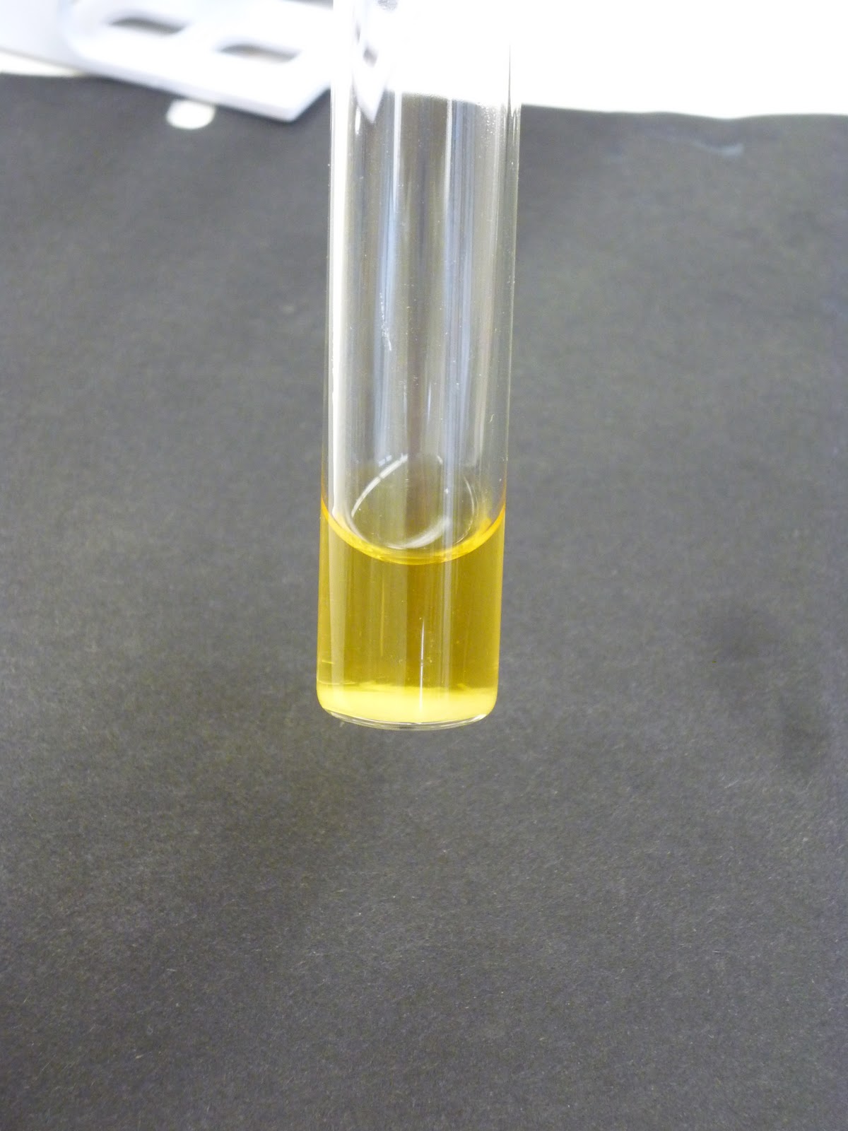
There was no change of color. The broth reminded yellow and did not turn pink. Thus my test was negative for Urea
Negative test for urea


Urea broth


OXIDASE
Purpose 
To determine if bacteria have cytochrome oxidase, a participant in electron transport during respiration. 
Materials
Oxidase reagent - N,N,N,N tetramethyl-p-phenylenediamine: the crushable ampule.
Sterile wood stick
piece of filter paper
Agar plate used for the Kirby-Bauer technique. 
Unknown Bacteria "K" in a new slant culture 

Procedure
I first prepared a new agar slant of my unknown bacteria using the procedure done in earlier tests.
I then incubated my new slant for 48 hours in 37 degrees C.
After incubation I used the crush-able ampule and I added a few drops of the oxidase reagent to the bacterial colony growing on my agar plate I used for the Kirby-Bauer technique.
I transferred some of the bacteria from my new slant onto the sterile wood stick and streaked the filter paper with it. A few drops of the oxidase reagent was applied.
Nothing happened which means my test was negative.

New agar slant to be inoculated

After incubation for 48 hours

Negative oxidase test


KIRBY-BAUER TECHNIQUE

Purpose 
To determine the sensitivity of a bacterium to several antibacterial medicines. 
Materials
Mueller-Hinton agar plate, 4 mm thick
antibiotic disk cartridges
forceps
95% ethanol in a beaker
ruler, millimeter
Unknown Bacteria "K" 


The Antibiotic Disks Used


Cell Wall
1. Penicillin
2. Vancomycin

Nucleic Acid
3. Novabiocin

Protein 
4. Tetracycline
5. Erythromycin
6. Chloramphenical
7. Neomycin
Procedure
I used aseptic technique to inoculate the Mueller-Hinton agar plate. I used an inoculating loop contaminated with the unknown bacteria to inoculate the entire surface of the agar with a closely spaced back-and-forth motion. I then inoculated the the agar surface a second and third time in different directions.

On the back side of my agar plate I labeled the areas where all 7 different cartridges would be placed evenly.  Before placing each different cartridge on the plate I dipped the forceps in alcohol, then burned the alcohol off.  I made sure to apply light pressure to the disks, using the forceps, to prevent them from fall off when the plate was to inverted for incubation.

I incubated it for 48 hours in 37 degrees C, making sure the plate was inverted.

After incubation I measured out the diameter in mm of the zone of growth inhibition for each disk. To do this I placed the ruler on the underside of the plate.

Agar plate I streaked with my unknown bacteria

The addition of the 7 different antibiotic disks

Here you can see how the different antibiotics affected my bacteria.NOTE: Number 1 disk is above the MS and the numbering continues counter-clockwise  with number 7 in the center.


My diameter results were as follows:
Cell Wall
1. Penicillin = NO inhibition took place --> Resistant
2. Vancomycin= 14 mm --> Sensitive

Nucleic Acid
3. Novabiocin= No inhibition took place --> Resistant

Protein 
4. Tetracycline= 25 mm --> Sensitive
5. Erythromycin= 11 mm --> Resistant
6. Chloramphenical= 34 mm --> Sensitive
7. Neomycin= 18 mm --> Sensitive

Monday, October 31, 2011

October 25-27

Tasks for this week:
EMB Agar
Mannitol Salt Agar
MacConkey

RESULTS: 
EMB: Dark purple, green metallic sheen
Mannitol Salt Agar: Mannitol non-fermenter
MacConkey Agar: Lactose-fermenter

EMB
Purpose:
To isolate and differentiate gram-negative enteric bacilli

Materials:
EMB agar plate
Unknown "K" bacteria

Procedure:
Label EMB plate correctly. (See previous blogs)
Use aseptice technique to inoculate the EMB agar plate as a streak plate.
Incubate the inoculated plate upside-sown for 48 hours at 37 degrees C.
See results


Notice the green metallic sheen.
MANNITOL SALT AGAR

Purpose:
To isolate bacteria base don their salt tolerance and differentiate among these isolates for mannitol fermentation.


Materials:
Mannitol salt agar plateUnknown "K" bacteria


Procedure:
Label Mannitol plate correctly. (See previous blogs)
Use aseptice technique to inoculate the Mannitol agar plate as a streak plate.
Incubate the inoculated plate upside-sown for 48 hours at 37 degrees C.

Look at Results
Mannitol non-fermenter. Note, my bacteria is on the right side and it did not grow.


MacConkey Agar

Purpose:
To detect and differentiate among gram-negative enteric bacilli, based on their ability to grow on the medium and to ferment lactose. 


Materials:
MacConkey agar plate
Unknown "K" bacteria


Procedure:
Label MacConkey plate correctly. (See previous blogs)
Use aseptice technique to inoculate the MacConkey agar plate as a streak plate.
Incubate the inoculated plate upside-sown for 48 hours at 37 degrees C.
See results
Notice the bubbles coming from the bacteria. This occurred after applying Hydrogen Peroxide to the bacteria. This again proves that my bacteria is filtrated.
Lactose-fermenter

Thursday, October 20, 2011

October 18 & 20

This week I completed various tests
  • Gelatin Hydrolysis test
  • Fermentation
  • Tripple Sugar Iron Agar test (TSIA)
  • Methyl Red Test
  • Litmus Milk Reaction
 RESULTS:  
  • Gelatin= negative 
  • Fermentation= Sugar and Acid were the end products. Gas was produced in both the glucose and lactose vials that were in the test tubes. My bacteria mostly used glucose and lactose. Not the sucrose, although there was some bacterial growth. 
  • TSIA = I observed a yellow slant/ yellow butt which = acid slant/ acid butt with gas. There was also a break in the agar. Which means that glucose and lactose &/ or sucrose were fermented.
  •  Methyl Red test = Positive test  for mixed-acid fermentation pathway. (my broth turned red)
  • VP Test= negative. My broth didn't change colors.
  • Litmus Milk reaction= results still unknown. Will not know until 7 days pass.
 Gelatin
Purpose:
To determine the ability of bacteria to hydrolyze gelatin. 
 
Materials needed:
Nutrient gelatin deep tube
unknown bacteria in slant

Procedure:
I labeled the test tube properly and used aseptic technique to inoculate the gelatin by stabbing the gelatin with an inoculating needle with bacteria from my unknown.

I stored the gelatin in the incubator at 37 degrees C for 48 hours then I placed it in the fridge for 15minutes. After removing the gelatin from the fridge I checked it for liquidation and the test came back negative because there was no liquidation.

Gelatin preinoculation


Gelatin after incubation and refrigeration. Note that liquidation did NOT occur. 


Fermentation of Carbohydrates

Purpose:
To determine the ability of some bacteria to ferment a particular carbohydrate. 


Materials:
3 tubes of phenol red- sugar broth with Durham tube
                                   The three sugars used were sucrose, lactose, and glucose. 
Unknown bacteria slant


Procedure:
Labeled the test tubes properly 
Used aseptic technique to inoculate each sugar broth with a loop full of unknown bacteria. 
I incubated the now contaminated broths in 37 degrees C for 48 hours although the instructions said for 24 hours. 
After incubation I checked each test tube for acid production or acid and gas production. 
My Lactose was very pale yellow. The glucose was a brighter shade of yellow and the sucrose stayed red. As seen in the picture below.


Lactose before being contaminated

Glucose before being contaminated

sucrose before being contaminated

Results. From Left to right: Lactose, Sucrose, Glucose.  
                                            Note the gas bubbles in both the lactose and glucose samples. 


 Triple Sugar Iron Agar Test (TSIA)
Purpose:
To differentiate among the gram-negative enteric bacilli as to their ability to ferment glucose, lactose, and sucrose and to produce H2S. 
 
Materials needed:
Triple sugar iron agar slant tube
unknown bacteria in slant
Inoculating needle with a straight wire

Procedure:
I labeled the TSI tube.
Used aseptic technique to remove a small amount of my unknown bacteria with an inoculating needle. I stabbed the bacteria into the TSI agar butt to about three-quarters deep. As I withdrew the needle I streaked in a zigzag pattern along the slant surface.
I incubated the tube at 37 degrees C for 24 hours and noted the changes that occurred.
My tube turned from red to completely yellow. Which means my slant is acidic and the butt had gas produced.
The Agar slant before being inoculated
After 24 hour incubation. Note the color change of the agar from red to yellow.

A better view of the color change that occurred


Methyl Red Test/ VP Test

Purpose:
To determine the ability of some bacteria to ferment glucose via mixed-acid fermentation.  


Materials:
Methyl red (MR-VP) broth tube
Unknown bacteria culture in slant
Barrett's reagent A
Barrett's reagent B
Disposable gloves
Methyl red in a dropper bottle 


Procedure:
Labeled the test tubes properly 
Used aseptic technique to inoculate the MR-VP broth with a loopful of my unknown bacteria. 
After incubating the broth at 37 degrees C for 48 hours I separated the broth into a second test tube (making sure there were equal amounts of contaminated broth in each tube.)
In the first tube I added 5 drops of methyl red. Gently swirled the tube to mix the broth culture and pH indicator.
After swirling I noted that a red color was formed. This redness yields a positive test for the mixed-acid fermentation pathway.


Broth after 48 hour incubation but before splitting it into two test tubes.

MR-VP positive test results
In the second tube I performed the VP test. 
I first put on the gloves before handling the Barrett's reagents.
VP negative test

I added 15 drops of Barrett's reagent A and 5 drops Barrett's reagent B.
After swirling the broth for a few minutes there was no color change which results in a negative test. 


Litmus Milk Reaction
Purpose:
To differentiate among bacteria as to their ability to utilize lactose, protein, and litmus in litmus milk. 
 
Materials needed:
Litmus milk tube
unknown bacteria in slant

Procedure:
I labeled the test tube properly and used aseptic technique to inoculate the litmus milk with a loopful of my unknown bacteria. 
I inoculated the tube at 37 degrees C for 48 hours. examined the tube, and placed it back in the incubator for another 4 days. 


Preinoculation

After 48 hours in the incubator